
-1024x226.jpg)
RXBio Translates Sequence to Science and Industry

The splicing of pre-mRNA is a process in which introns are removed and exons are spliced together to form mature mRNA under the catalysis of the spliceosome. It is a necessary step in gene expression in higher eukaryotes and also one of the key mechanisms for generating the diversity of protein molecules.
The entire splicing process is completed in the nucleus and is under fine and strict regulation. Mutations at splicing sites will inevitably lead to changes in splicing patterns. The correct sequence and position of splicing sites are both necessary conditions to ensure the normal splicing of RNA.
The splicing process requires three important splicing signals, namely the 5’ splicing site (donor site), the 3’ splicing site (acceptor site), and the branch site (near the 3’ end of the intron). The 5’ splicing site generally consists of GU followed by several bases that are not particularly conserved. The 3’ splicing site region contains multiple conserved elements, which are, in sequence, the branch point, the polypyrimidine tract, and the AG at the 3’ end of the intron. In addition, the splicing process also requires the assistance of some other cis-regulatory elements. According to their locations and functions, they can be divided into four categories: exon splicing enhancer (ESE), exon splicing silencer (ESS), intron splicing enhancer (ISE), and intron splicing silencer (ISS). Generally, these auxiliary regulatory elements promote or inhibit the recognition of splicing sites by recruiting some trans-acting factors, that is, splicing regulatory proteins, and can regulate the splicing process by regulating the assembly of the spliceosome. The currently known splicing regulatory proteins are mainly from the serine/arginine-rich protein (SR protein) family and the heterogeneous nuclear ribonucleoprotein (hnRNP) family. Generally, SR proteins bind to purine-rich ESE and ISE, promoting the binding of U1 snRNP to the 5’SS, the binding of U2AF to the 3’SS, and the assembly of the U4/U6-U5 snRNP trimer onto the spliceosome, thereby enhancing the splicing efficiency. Members of the hnRNP family usually bind to ESS and ISS to inhibit the recognition and utilization of splicing sites. Only through the complex interactions between cis-acting elements and trans-acting factors can the correct splicing process be achieved.
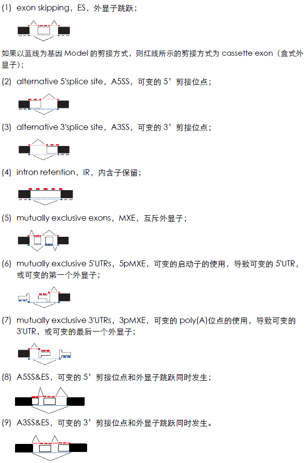
Alternative splicing patterns are mainly classified into exon skipping (ES), alternative 5’ splice site (A5SS), alternative 3’ splice site (A3SS), intron retention (IntronR), mutually exclusive exons (MXE), mutually exclusive 5’ UTRs (5pMXE), mutually exclusive 3’ UTRs (3pMXE) (Wang, Sandberg et al. 2008). Besides these, there are also cassette exon, A3SS&ES and A5SS&ES.
In the analysis of alternative splicing events, one of the multiple annotated transcripts of each gene needs to be selected as the gene model, that is, the reference transcript, and then the transcripts with alternative splicing are analyzed relative to the gene model. The schematic diagram of alternative splicing events is as follows (the red line corresponds to the inclusive/extended isoform, the long transcript, and the blue line corresponds to the exclusive isoform, the short transcript).
If changes in certain nucleotides affect splicing signals and functional elements, the splicing pattern of genes will change, thus leading to the occurrence of certain diseases. On the other hand, by changing the splicing pattern of genes, the clinical phenotypes of some diseases can also be modified. With the application and development of antisense oligonucleotide drugs in the clinic, the mini-gene can identify specific nucleotide sequences that affect the splicing pattern, thus providing an effective method for finding drug treatment targets. In addition, the mini-gene can screen some targeted therapeutic drugs by studying the impact of certain compounds on the gene splicing pattern, and it can be used for drug screening for spinal muscular atrophy, muscular dystrophy, familial dysautonomia and other genetic diseases.
Among them, Novartis screened approximately 1.4 × 10⁶ compounds through the Minigene reporter technology and obtained small molecule compounds that can stabilize the structures of SMN2 pre-mRNA and U1 snRNP (a key component of the spliceosome), thereby increasing the proportion of correct splicing of SMN2 mRNA.
· Client Provision: Genes to be tested of interest, RNA-seq data, iRIP/CLIP-seq data
· Result Delivery:
1) Plasmids constructed by minigene
2) Schematic diagram of minigene sequence design
3) Experimental report (including detailed experimental steps, manufacturer and catalog number information of main instruments and reagents)
4) English materials and methods